Dental curricula increasingly rely on high‑resolution printable tooth anatomy models to bridge the gap between textbook diagrams and hands‑on experience. By converting 3D scans into PDF or STL files that students can print at home, educators provide an inexpensive, repeatable resource that reinforces morphology, pathology, and restorative concepts.
Why Printable Models Enhance Learning
Traditional cadaveric specimens are costly, limited in availability, and often unsuitable for repeated dissection. Printable models offer three distinct advantages:
- Portability: A single file can be accessed on any computer, allowing learners to study anywhere with a printer.
- Customization: Instructors can annotate layers, highlight anomalies, or isolate structures such as the pulp chamber, enamel rods, or periodontal ligament.
- Scalability: One digital file yields unlimited physical copies, supporting large cohorts without additional material expense.
Step‑by‑Step Guide to Generating a Printable Tooth Model
Creating a usable model involves three technical stages, each achievable with free or low‑cost software.
- Data acquisition: Obtain a high‑resolution CBCT or micro‑CT scan of the target tooth. Export the dataset in DICOM format.
- Segmentation: Use open‑source tools like 3D Slicer to isolate enamel, dentin, pulp, and root structures. Apply thresholding to differentiate tissue densities.
- Export and preparation: Convert the segmented volumes to STL files, then clean meshes in MeshLab to remove non‑manifold edges. Finally, generate a printable PDF by flattening the 3D surface into a 2D pattern for paper‑based models or upload the STL to a slicer for resin printing.
Best Practices for Dental Students
To maximize the educational return on printable models, adopt the following workflow:
- Pre‑print review: Examine the digital model on a tablet, noting critical landmarks such as the cementoenamel junction and occlusal pits.
- Print in appropriate scale: For detailed anatomy, a 1:1 size is ideal; for comparative studies, a 1:2 scale may reduce material use while preserving feature visibility.
- Post‑print annotation: Use colored markers to label roots, fissures, and carious lesions. This tactile interaction reinforces spatial memory.
- Integrate with case studies: Pair each model with a clinical vignette—e.g., a molar with proximal caries—to contextualize the anatomy.
Implications for Curriculum Design
Institutions that embed printable models into their courses report higher quiz scores in morphology sections and greater student confidence during clinic rotations. The modular nature of digital files also allows rapid updates when new research modifies anatomical definitions or when novel pathologies emerge.
Looking Ahead: Hybrid Learning and Emerging Technologies
While paper‑based prints remain valuable for tactile learners, the rise of affordable desktop resin printers is shifting the focus toward fully three‑dimensional replicas. Future iterations may incorporate augmented reality overlays, enabling students to visualize internal structures without dissection. As file repositories expand, collaborative platforms will let educators share annotated models globally, standardizing dental education across regions.
Free May Printable Calendar: Plan Your Month! - Printables For Everyone
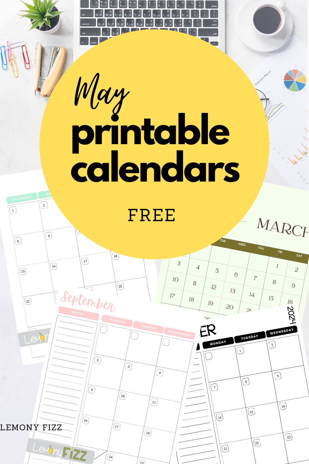
Free May Printable Calendar: Plan Your Month! - Printables for Everyone
Free Printable 2025 Calendar Templates All 12 Months – Artofit

Free printable 2025 calendar templates all 12 months – Artofit
Printable Alphabet Letters For Little Girls

Printable Alphabet Letters For Little Girls
Free Printable Girly
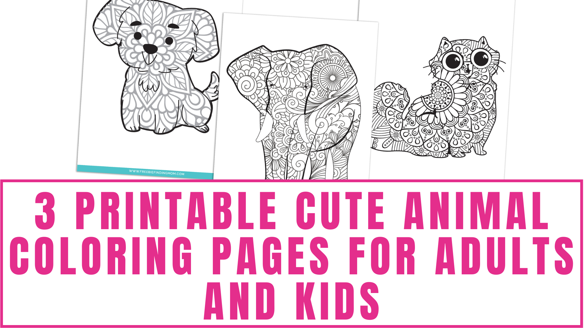
Free Printable Girly
Coloring Worksheet Page. Educational Printable Coloring Worksheet

Coloring worksheet page. Educational printable coloring worksheet ...